Introduction:
The Importance of Vision Our eyes are our primary sensory organs, allowing us to perceive the world in all its splendor. Vision shapes our experiences, influences our decisions, and connects us to our surroundings. Imagine a life without the ability to see—no vibrant sunsets, no smiling faces, no breathtaking landscapes. Vision is not just a biological function; it’s a gateway to understanding, wonder, and connection.
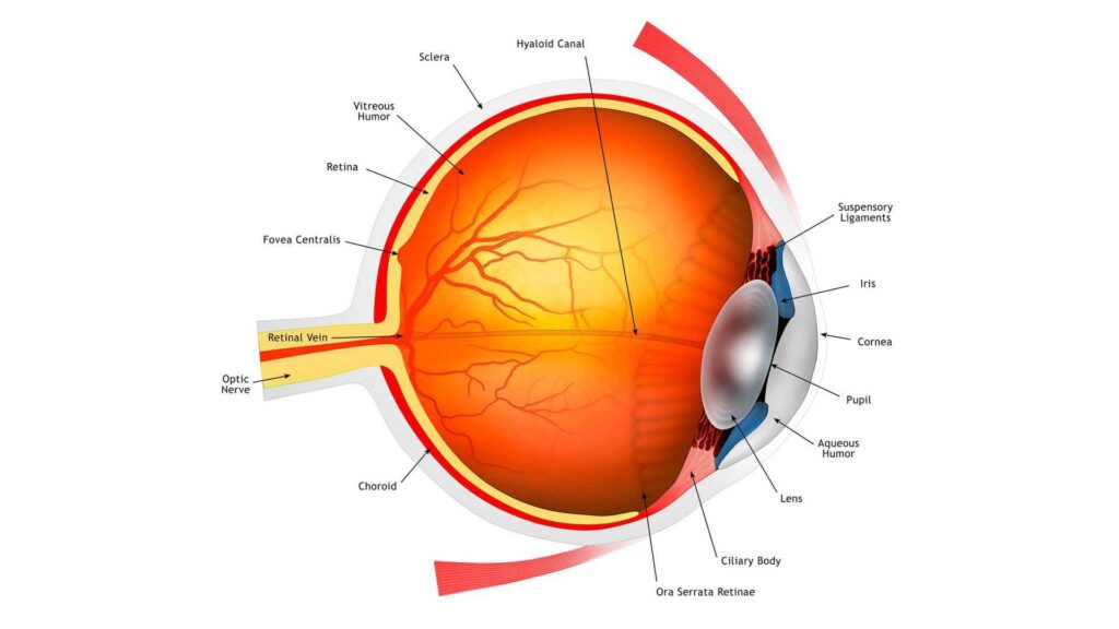
The Role of the Human Eye: The human eye is an intricate masterpiece of evolution. Its primary function is to capture light and convert it into electrical signals that our brain interprets as visual information. Here’s a brief overview of how it works:

- Light Entry: Light enters the eye through the transparent cornea—the eye’s protective outer layer. The cornea bends and focuses the incoming light.
- Adjustable Aperture: The pupil, located in the center of the colored iris, acts as an adjustable aperture. It regulates the amount of light entering the eye. In bright conditions, the pupil constricts; in dim light, it dilates.
- Lens and Focus: Behind the pupil lies the lens. This flexible structure fine-tunes focus by changing its shape. It adjusts to focus on objects at varying distances.
- Retina and Photoreceptors: At the back of the eye, the retina awaits. It contains millions of specialized cells called photoreceptors—rods and cones. Rods are sensitive to low light and help us see in dim conditions, while cones allow us to perceive color and detail.
- Signal Transmission: When light strikes the retina, photoreceptors convert it into electrical signals. These signals travel via the optic nerve to the brain’s visual cortex, where they’re transformed into the images we perceive.
Anatomy of the Eye: Cornea and Pupil
Cornea: The Transparent Window
- The cornea is the eye’s clear, curved outermost layer.
- It acts like a transparent window, allowing light to enter the eye.
- Light rays bend as they pass through the cornea, focusing them onto the lens.
Pupil: The Adjustable Aperture
- The pupil is the dark circular opening in the center of the iris (the colored part of the eye).
- It functions like an adjustable aperture in a camera.
- In bright light, the pupil constricts (becomes smaller) to limit the amount of light entering the eye.
- In dim light, the pupil dilates (expands) to allow more light in.
Iris and the lens:
Iris: Regulating Light
- The iris, that colorful part of the eye, plays a crucial role in controlling the amount of light entering.
- In bright conditions, the iris constricts the pupil, making it smaller to limit light.
- In dim light, the iris relaxes, widening the pupil to allow more light in.
Lens: Focusing Light
- Directly behind the pupil sits the lens.
- The lens focuses incoming light toward the back of the eye.
- It adapts its shape to help us focus on objects at varying distances.
Retina and its photoreceptors:
Retina:
- The retina is a thin, light-sensitive layer located at the back of the eye.
- It plays a crucial role in vision by capturing light and converting it into electrical signals.
- These signals are then transmitted to the brain via the optic nerve for further processing.
Photoreceptors:
- Photoreceptors are specialized cells within the retina that detect light.
- There are two main types of photoreceptors:
- Rods: These cylindrical-shaped cells are responsible for night vision (scotopic vision).
- Rods contain a protein called rhodopsin.
- They do not perceive color but allow us to see in low-light conditions.
- Cones: These conical-shaped cells enable color vision and function best in bright light (photopic vision).
- Cones contain proteins called photopsins (cone opsins).
- There are three types of cones: blue, red, and green, each sensitive to specific wavelengths.
- Cones dominate the central part of the retina (macula) and help us see fine details and colors.
- Rods: These cylindrical-shaped cells are responsible for night vision (scotopic vision).
Importance:
- Photoreceptors are essential for our visual experience:
- Night Vision: Rods allow us to see in dim light, which is crucial for activities like driving at night.
- Color Vision: Cones help us distinguish different colors and perceive bright light.
Vision Processing
Retina and Photoreceptors:
- When light enters the eye, it passes through the cornea and lens, forming an inverted image on the retina.
- The retina contains specialized cells called photoreceptors: rods and cones.
- These photoreceptors convert light into electrical signals.
Optic Nerve:
- The optic nerve is a bundle of nerve fibers originating from the photoreceptors in the retina.
- These fibers transmit the electrical impulses generated by photoreceptors.
- The optic nerve carries these signals to the brain for interpretation.
Visual Processing:
- The information from the retina is relayed via the optic nerve to the lateral geniculate nucleus in the thalamus.
- From there, it reaches the primary visual cortex in the occipital lobe at the back of the brain.
- The primary visual cortex processes the signals, allowing us to perceive the visual world.
Visual Cortex
Location and Function:
- The visual cortex is located in the occipital lobe of the brain.
- Its primary role is to interpret and process visual input received from the eyes.
Processing Visual Signals:
- Sensory input from the eyes travels through the lateral geniculate nucleus in the thalamus.
- From there, it reaches the visual cortex.
- The primary visual cortex (V1), also known as Brodmann area 17, receives this input.
- Neurons in V1 fire action potentials when visual stimuli appear within their receptive field.
- Receptive fields are specific regions of the visual field that trigger neural responses.
- Neurons in V1 exhibit retinotopic organization, meaning neighboring cells correspond to adjacent portions of the visual field.
Complex Tuning:
- Neurons in higher visual areas (such as the inferior temporal cortex) have complex tuning.
- They respond selectively to specific features (e.g., faces) within their receptive fields.
- Contextual modulation also plays a role, in shaping our visual experiences.
Accommodation and Focus
Accommodation:
- Definition: Accommodation refers to the eyes’ ability to see objects clearly at different distances by adjusting the optical power.
- Structures Involved:
- Ciliary Muscle: A smooth ring-shaped muscle in the middle of the eye.
- Holds the lens via suspensory ligaments.
- Adjusts the lens shape during accommodation.
- Lens: A transparent, biconvex structure.
- Changes shape to focus light onto the retina.
- Becomes rounder (more convex) for near objects and flatter for distant objects.
- Pupil: The black opening in the center of the eye.
- Constricts to prevent scattered light from reaching the retina.
- Ciliary Muscle: A smooth ring-shaped muscle in the middle of the eye.
Accommodation Reflex:
- When we shift focus from a distant object to a nearby one:
- Ciliary Muscles Contract: These muscles tighten.
- Lens Shape Changes: The lens becomes more spherical.
- Increased refractive power allows sharper bending of light rays.
- Result: Clear vision for objects at varying distances.
Common Eye Conditions
Myopia (Nearsightedness):
- Definition: Myopia occurs when distant objects appear blurry.
- Cause: The eyeball is too long from front to back.
- Focus: Light focuses in front of the retina.
- Vision: Nearby objects look clearer, but distant ones are unclear.
- Prevalence: About 40% of Americans have myopia.
Hyperopia (Farsightedness):
- Definition: Hyperopia causes close objects to appear blurry.
- Cause: The eyeball is too short.
- Focus: Light focuses behind the retina.
- Vision: Distant objects are clearer, but close ones blur.
- Prevalence: Only 5-10% of Americans experience hyperopia.
Cataracts and glaucoma
Cataracts:
- Definition: Cataracts involve the clouding of the eye’s natural lens, which sits behind the iris and pupil.
- Cause: Aging, UV exposure, or other factors lead to protein changes in the lens, causing opacity.
- Symptoms: Gradual vision decline, hazy or cloudy vision, glare sensitivity.
- Treatment: Surgical removal and replacement with an artificial lens.
Glaucoma:
- Cause: Glaucoma results from increased fluid pressure inside the eye (aqueous humor).
- Types: Open-angle (gradual) and closed-angle (sudden).
- Symptoms: Peripheral vision loss (open-angle), intense eye pain (closed-angle).
- Treatment: Eye drops, laser surgery, or other interventions to manage pressure.
Fun Facts and Curiosities
Inverted Retinal Image:
- When light enters your eye, it forms an image on the retina at the back of your eyeball.
- Surprisingly, this image is reversed and upside down!
- Imagine looking through a keyhole to spy on someone in a room. You’d need to move your head in the opposite direction to see the entire room. Similarly, our pupils act like keyholes, allowing us to see larger objects by reversing the image.
- The brain then reorients the image, so you perceive things “right side up” despite the initial inversion. This adaptation gives us tremendous peripheral vision and the ability to view objects much larger than a few millimeters.
576-Megapixel Resolution:
- Can the human eye see in 8K? Well, sort of!
- Someone did the math, assuming 20/20 vision, and estimated that our eyes can process an astounding 576 megapixels.
- That’s roughly 576 million individual pixels! So, in terms of resolution, our eyes surpass what an 8K TV offers.
- However, keep in mind that our visual system is more complex than just pixel count. Factors like color perception, dynamic range, and motion sensitivity also play a role in how we perceive the world.
Conclusion: The Marvel of Our Eyes
Our eyes are truly remarkable organs, combining intricate design with astonishing functionality. Here’s a recap of their complexity and importance:
Biological Cameras:
- The eye acts as a biological camera, capturing light and transforming it into electrical signals that our brain interprets as visual information.
- Its lens, cornea, and iris work together to focus light onto the retina, where millions of photoreceptor cells await.
Retina and Photoreceptors:
- The retina, located at the back of the eye, contains specialized cells called photoreceptors: rods and cones.
- Rods allow us to see in low light conditions, while cones provide color vision.
- These cells convert light into electrical impulses, which travel along the optic nerve to the brain.
Incredible Adaptations:
- Our eyes adapt to varying light levels, adjusting the size of our pupils and the sensitivity of our retinal cells.
- Blink reflexes protect our eyes from debris, and tear production keeps them moist and clear.
Visual Perception:
- The brain processes visual input, reconstructing the world around us.
- It corrects the inverted retinal image, allowing us to perceive objects right side up.
- Our eyes provide peripheral vision, depth perception, and motion detection.
576-Megapixel Resolution:
- While not exactly like a digital camera, our eyes process an estimated 576 megapixels of visual information.
- This resolution surpasses even the most advanced screens, emphasizing the eye’s incredible detail.
Daily Dependence:
- Our lives revolve around visual experiences: reading, recognizing faces, appreciating art, and navigating our surroundings.
- The eye’s role extends beyond vision—it also influences our emotions and well-being.
FAQ’s
What are the main parts of the human eye?
The main parts of the human eye include the cornea, lens, iris, pupil, retina, optic nerve, and vitreous humor. Each part plays a crucial role in the process of vision.
How does light enter the eye?
Light enters the eye through the cornea, which is the transparent, dome-shaped surface that covers the front of the eye. The cornea helps to focus the incoming light.
What is the function of the pupil?
The pupil is the opening in the center of the iris that controls the amount of light entering the eye. It dilates (expands) in low light to allow more light in and constricts (shrinks) in bright light to reduce light entry.
Why is regular eye care important?
Regular eye care is important to detect and treat vision problems early, maintain overall eye health, and prevent conditions that could lead to vision loss. Regular check-ups with an eye care professional can help monitor changes in vision and eye health.


